AI skin diagnostics are image-based tools that estimate visible skin concerns from a photograph and return severity scores, zone-by-zone breakdowns, and product suggestions. They do not produce a medical diagnosis. Typical outputs include hydration, texture, tone, acne level, pore size, redness, and a composite skin-age metric. Accuracy depends on capture conditions, camera quality, and the diversity of the training data the model was built on.
Table of Contents
- How do AI skin diagnostics actually work?
- What does the user experience actually look like?
- How accurate are these tools, and where do they fall short?
- How should you interpret results safely?
- How do you choose a trustworthy tool in the UK?
- Where do these tools genuinely add value?
- Key takeaways
- A practical perspective on using AI diagnostics when buying a home device
- Useful sources and further reading
How do AI skin diagnostics actually work?
At the core of most consumer tools is computer vision: the system breaks a selfie into a pixel map and compares it against large, labelled image datasets to detect patterns associated with acne, pigmentation, fine lines, and pores. The model assigns probability scores to each detected concern, then a recommendation engine maps those scores to suggested products or routines. Multimodal systems that also collect medical history, declared skin type, and location tend to produce more contextually grounded suggestions than image-only tools.
Not all systems use the same imaging approach. Understanding the differences helps you evaluate marketing claims such as “clinically validated” or “deep skin analysis.”
- Computer vision (RGB camera): Analyses surface-level pixel patterns. Standard in smartphone apps. Cannot detect subsurface structures.
- Multispectral imaging: Uses UV, cross-polarised, and infrared light to reveal subsurface damage, pigmentation below the skin surface, and bacterial markers (porphyrins). Found in professional clinic systems and some advanced home devices.
- Spectroscopy: Measures light absorption at specific wavelengths to infer biochemical properties. Rare in consumer devices; more common in research and clinical settings.
| Technology | What it measures | Typical setting |
|---|---|---|
| Computer vision (RGB) | Surface texture, tone, visible lesions | Smartphone apps, web tools |
| Multispectral imaging | Subsurface pigment, UV damage, porphyrins | Clinic systems, pro-grade devices |
| Spectroscopy | Biochemical skin properties via light absorption | Research, specialist clinics |
Claims of “deep skin” measurement from a single RGB selfie should be treated with scepticism. Professional systems use multi-angle capture with UV and cross-polarised lighting to produce clinically different outputs that a smartphone camera simply cannot replicate. For a closer look at how device microprocessors shape these capabilities, the hardware differences matter considerably.
What does the user experience actually look like?
The typical workflow is faster and simpler than most people expect.
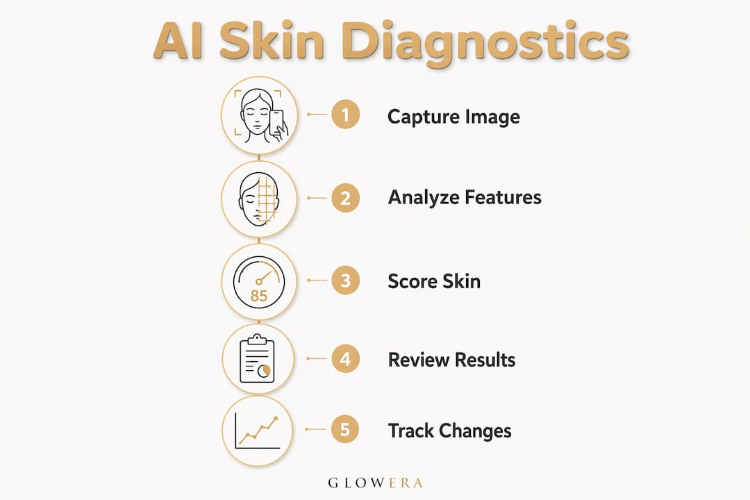
- Open the app or web tool. Some platforms are browser-based; others require a dedicated app.
- Prepare your skin. Remove makeup, cleanse, and wait a few minutes for skin to settle.
- Set up your lighting. Position yourself facing a natural or diffuse white light source, avoiding harsh side-lighting or shadows.
- Take the selfie. Hold the camera at eye level, maintain a neutral expression, and keep the lens clean.
- Wait for processing. Most consumer tools return results in seconds.
- Read the report. Review zone-by-zone scores for hydration, texture, tone, acne, and pores. Note any flagged concerns.
- Schedule follow-up scans. Consistent repeat scans, taken under the same conditions, are what generate reliable trend data.
Pro Tip: The single variable that most affects trend reliability is lighting consistency. Use the same spot, at the same time of day, every scan. Skin changes are subtle; inconsistent lighting will mask them or create false positives.
For practical guidance on building a tracking habit, Glowera’s guide on tracking skincare device progress covers the capture and logging workflow in detail.
How accurate are these tools, and where do they fall short?
Independent clinical studies report correlations with physician assessments often above 80% for specific metrics, with test-retest reliability cited at 95% in industry summaries. A systematic review of AI skin diagnostics in primary care settings found sensitivity ranging widely across studies across studies, with accuracy varying considerably depending on the algorithm and task depending on the algorithm and task. For melanoma detection specifically, a meta-analysis reported pooled sensitivity of approximately 0.86 and specificity of approximately 0.88 in controlled settings.
Key benchmark: Test-retest reliability and physician correlation are reported to be high in controlled laboratory or clinical settings, though real-world performance may be lower. These figures apply to controlled capture conditions — real-world performance is lower.
The main sources of error in consumer tools:
- Lighting: Mixed or directional light creates shadows that the model may read as texture or pigmentation changes.
- Angle: Even a slight tilt alters which zones the model maps to which facial regions.
- Camera quality and lens cleanliness: Low-resolution sensors and smudged lenses introduce noise that degrades classification.
- Image compression: Heavily compressed JPEGs lose fine detail that the model relies on.
Training-data bias is a documented and serious concern. A 2025 study found that 89.8% of generated dermatological images depicted light skin tones, with measurable accuracy drops on Fitzpatrick IV–VI phototypes. Models trained predominantly on lighter skin tones perform less reliably on darker complexions, and conditions such as melanoma, eczema, and rosacea present differently on deeper skin. Always check whether a tool discloses its Fitzpatrick coverage before relying on its scores.
Pro Tip: To reduce false positives, take three scans in the same session and compare them. If a concern appears in all three, it is more likely genuine. A concern that appears in only one scan is more likely a capture artefact.
For context on reading clinical validation claims on device packaging, understanding what “validated” actually means in practice is worth the extra five minutes.
How should you interpret results safely?
Consumer AI tools are orientation and tracking instruments, not diagnostic devices. Dermatology experts consistently emphasise AI’s role as a complement to clinical assessment rather than a replacement. A tool that claims to deliver a definitive medical diagnosis exceeds its safe design boundary.
Practically, this means treating scores as directional signals. A rising texture score over eight weeks of LED therapy is useful feedback. A single high severity score for a lesion is not a diagnosis.
Seek a dermatologist promptly if you notice:
- A new or changing lesion that persists beyond four weeks
- A red patch, ulcer, or wound that does not heal
- Any mole that changes in size, shape, or colour
- Symptoms accompanied by pain, bleeding, or crusting
On privacy: before using any tool, check its data policy for three things. First, whether your images are stored and for how long. Second, whether your scans can be used to retrain the model without your explicit consent. Third, the data jurisdiction, particularly relevant for UK users under UK GDPR.
Pro Tip: If you have a high-stakes concern, use an AI scan to document the current state with a timestamped image, then take that report to a dermatologist or in-clinic imaging session. The AI output gives the clinician a useful visual reference point, even if it carries no diagnostic weight.
For guidance on combining AI tracking with personalised skincare and clinical treatment pathways, a med-spa consultation can help contextualise what your scores actually mean.
How do you choose a trustworthy tool in the UK?
Not all tools are equal, and marketing language can obscure meaningful differences. Use this checklist when evaluating any AI skin diagnostic tool or device:
- Declared imaging method: Does the tool state whether it uses RGB, multispectral, or spectroscopy? Vague claims about “advanced imaging” are a warning sign.
- Clinical validation: Are there third-party peer-reviewed studies, not just internal accuracy claims?
- Training-data diversity: Does the tool disclose Fitzpatrick I–VI coverage? If not, assume the model may underperform on darker skin tones.
- Transparency and explainability: Does the report explain what the model detected and why, or does it only output a score?
- Privacy and data retention: Can you opt out of having your images used for model training? Where is data stored?
- Regulatory status: In the UK, tools making medical diagnostic claims must meet UKCA or CE marking requirements as medical devices. Cosmetic and wellness tools operate under a different, lighter framework. Know which category your tool falls into.
- Progress tracking: Does the tool store scan history and show trends over time, or does it only return a one-off result?
When to prefer multispectral or professional-grade devices: if you are tracking post-treatment recovery, monitoring a suspicious lesion, or using the tool to guide high-investment device purchases, the additional data from a multispectral system justifies the cost. For routine habit support and product testing, a well-validated smartphone app is sufficient.
Be cautious of free consumer tools whose recommendation engines map concern scores directly to a proprietary product catalogue. The scan may be accurate; the recommendation is a commercial output, not a clinical prescription.
Where do these tools genuinely add value?
AI skin diagnostics are most useful when the goal is measurable, visual, and tracked over time.
- Progress tracking for at-home treatments: Scanning every two to four weeks during an LED or microcurrent course gives you objective texture and tone data rather than subjective impression. Eight to twelve weeks is a realistic window to see meaningful change in scores.
- Pre-treatment baseline: A scan before starting a new device or routine creates a reference point. Without it, you cannot distinguish genuine improvement from natural skin variation.
- Routine optimisation and product testing: Comparing scores before and after introducing a new serum or device helps isolate what is actually working.
- Triage for suspicious changes: A scan can document a concern with a timestamped image before a dermatologist appointment, giving the clinician useful visual context.
These tools are less useful for diagnosing rashes or inflammatory conditions, assessing internal hydration (which RGB cameras cannot measure), or evaluating anything that requires dermoscopy or biopsy. Skin cancer assessment from a consumer selfie tool carries real risk of both false reassurance and unnecessary alarm.
If you are pairing AI diagnostics with a professional device, Glowera’s range of K-beauty tech devices includes clinic-validated hardware designed for exactly this kind of tracked, at-home treatment workflow.
Key takeaways
AI skin diagnostics are useful orientation tools when used with consistent capture conditions and realistic expectations, but they do not replace clinical assessment for anything medically significant.
| Point | Details |
|---|---|
| Outputs are estimates, not diagnoses | Tools return severity scores and skin-age metrics; they do not diagnose skin conditions. |
| Capture consistency is critical | Lighting, angle, and lens cleanliness affect scores more than small skin changes. |
| Training-data bias is real | A 2025 study found 89.8% of dermatological training images showed light skin tones; check Fitzpatrick I–VI disclosure. |
| Validate before you trust | Look for third-party clinical studies, Fitzpatrick coverage disclosure, and a clear privacy policy. |
| See a clinician for red flags | Persistent lesions, non-healing wounds, or changing moles require dermatologist review, not a rescan. |
A practical perspective on using AI diagnostics when buying a home device
The most common mistake buyers make is treating a single AI scan as a verdict. It is not. What these tools do well is give you a repeatable, visual baseline, and that is genuinely valuable when you are investing in a professional-grade device and want to know whether it is working.
For high-stakes needs, a multispectral or clinic-validated system will always outperform a smartphone app. The subsurface data it captures simply cannot be replicated by an RGB camera, regardless of how sophisticated the algorithm is. For everyday tracking and habit support, a well-validated app with declared Fitzpatrick I–VI coverage and a transparent privacy policy is entirely fit for purpose.
The advice worth following: run a baseline scan before you start any new device or routine, repeat it under identical conditions every four weeks, and bring the trend data to a dermatologist if anything looks unusual. AI gives you the documentation; the clinician provides the interpretation.
Useful sources and further reading
The following sources back the claims in this guide and are worth consulting directly for deeper technical detail or regulatory context.
- PMC systematic review on AI skin diagnostics in primary care: Covers 15 studies (2019–2022) on AI diagnostic accuracy for skin conditions, with sensitivity ranging widely across studies. Useful for understanding real-world performance variation.
- Meta-analysis: AI vs. clinicians for skin conditions: Reports pooled sensitivity of approximately 0.86 and specificity of approximately 0.88 for melanoma detection. Backs the accuracy benchmarks cited in this guide.
- MDPI review: AI methods in dermatology: Comprehensive overview of deep learning architectures, regulatory frameworks, and ethical considerations in AI dermatology.
- Expert commentary on AI in cosmetic dermatology: Backs the orientation-versus-diagnosis framing and the expert consensus that AI complements rather than replaces clinical assessment.
- Beauty Tech Review: smart mirrors and AI skin scanners: Source for the 2025 training-data bias finding (89.8% light skin tones) and the explanation of multispectral versus RGB imaging.
This article is general consumer information, not medical advice. For any skin concern that may require diagnosis or treatment, consult a qualified dermatologist or healthcare professional.